Light Sensitivity in Corneal Neuralgia
Structured explanations of photophobia, mechanisms, and patient observations. Designed for both reading and audio accessibility.
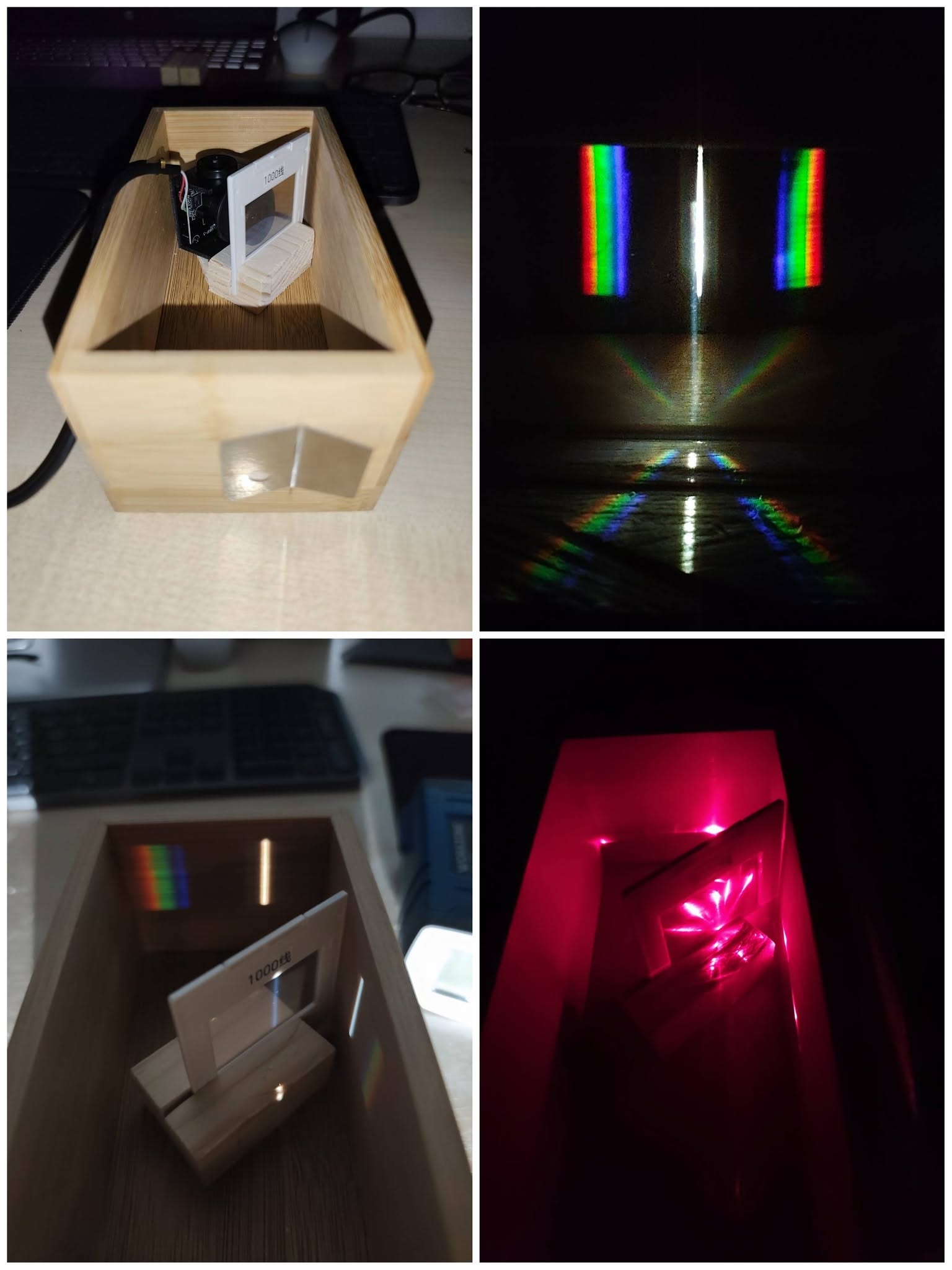
Hello my fellow sufferers. I'm happy to announce what I've been planning for months. I've built a mini spectrometer, to analyse light spectrum, and at the same time reading about light sensitivity to try to figure it out, since it's been playing a major role in my CN, and I'm sure it's been a big problem for many of you. I am managing light sensitivity better than I was a year ago (especially given that my dry eye has independently progressed all the way), but I cannot say I really cured myself. I also understand the mechanism of the light sensitivity more and it's quite interesting.
I hope these upcoming posts will help many of you. At bare minimum - to help you understand more, and get new ideas on how to improve yours. Because I have a lot to say and one post would be too long, I will divide what I have to say in 3-5 posts. Also I will try to make it short and concise. It's ironic, that this post is meant for people that cannot or have a hard time reading it 🙁
In this post I want to start by basics of blue light, so this one will be short. But in the upcoming posts I want to discuss about the pain mechanisms, why some of you have pain with screens but not sunlight and vice versa, and last but not least - discuss about different screen types, and how protection with these works in practice, with my amateur spectrometer.
But first let me just say, some of the things I will say could be wrong, (I hope not), because I really don't want to mislead anyone. I would be more than happy to get corrected by some nerds who got into this even more than me. After all, the point of these posts is to help us all. I also wish to discuss with you about your light sensitivity and symptoms, so we can learn even more. That said, feel free to tell about your own light problems in the comments.
Introduction: Blue Light Basics
Light comes in different colors, and each color has a wavelength. Shorter wavelength means more energy per particle of light. Almost every modern light source (phones, tablets, computers, TVs, projectors, home lighting, streetlights, some car headlights…), has this blue spike (see the 3rd picture), some more, some less. The tricky part is: the light doesn’t have to look blue for it to be there. A screen or lamp that looks white can still hide a big blue peak and cause major pain.
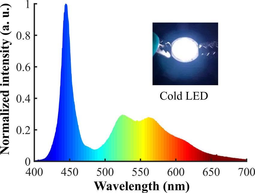
Why? Because “white” isn’t a single color - it’s a mix of many colors together. Sunlight spreads them out evenly, like a smooth rainbow. But LEDs usually make white by blasting a blue core and then adding yellow, which tricks your eye into seeing “white.” That’s why the blue spike shows up on the graph. Examples:
Blue object looks blue because it reflects mostly blue light and absorbs other colors (red, green, etc.).
White object: it looks white because it reflects almost all colors, so even though it is white, it does NOT mean there is no blue color, but rather the blue is hidden in a mix of other colors, roughly equally. The relative intensity is just less, than let's say, in a blue object.
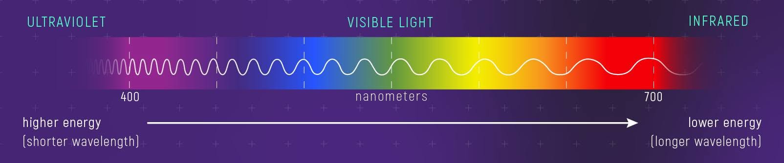
The visible spectrum goes from about 380 nm (violet/blue, high energy) to 700 nm (red, lower energy), (see the 2nd picture). Shorter waves means more energy which means more stimulation for sensitive eyes and nerves. That’s why blue feels harsher than red.
Next post will be about light induced pain mechanism, and it will be longer and detailed.