Light Sensitivity in Corneal Neuralgia
Structured explanations of photophobia, mechanisms, and patient observations. Designed for both reading and audio accessibility.
In this post I want to discuss different pain mechanisms triggered by light. It's very important topic to understand and in the last 4th section I'll even explain how you could maybe help your light sensitivity if it is centrally modulated. I assume many of you experienced pain by looking at screen whether it's from the phone, computer, TV... Doctors tell you to blink more, so you blink and blink, but the pain is still there, even when Schirmer's and TBUT are normal. This happened to me (still happens, but my eye is now also severely dry, so it's harder to objectively differentiate between dry eye and photophobia).
1. CHRONIC PROBLEMS OF BLUE LIGHT
What is causing this pain? Why not from normal ambient light, which is in fact far more bright than a little screen? A major part of the answer lies in blue wavelengths. Chronic exposure to blue light, in vivo and in vitro, is known to cause:
- oxidative stress
- inflammation
- cell death
- impaired healing
Blue light increases the production of reactive oxygen species (ROS) in corneal epithelial cells, damaging the ocular surface. Studies also show that chronic exposure to blue light inhibits the migration and repair capabilities of corneal epithelial cells, slowing down wound healing. High exposure can cause corneal epithelial thinning and increased cellular vacuolization.
All of this however happened in experimental studies under high intensity and prolonged exposure [1]. For example there's a study about mice and human corneal epithelial cells where the intensity used was 2.5 W/m2, and exposure was done for 4, 12, and 24 hours. To put that into perspective, phone's worst case scenario delivers 1-2 W/m2 of blue light. That's just 1.25 - 2.5x less than in the experiment. But it's also assuming phone being on full brightness, 15 cm (6 inches) away from your eyes. Distance here is a big factor, cut the distance in half, and you get 4x more energy intensity (although that's not exactly true since phone is not a point source), so the factor would be more like ~2x more instead. This still means if you have your phone really up close you can surpass the intensity in the study. However don't be alarmed yet, because blinking and natural healing of the tissue, lowers the chances of these problems in practice.
At the same time the paper does not specify a strict “no-effect” threshold below 2.5W/m2 (as that was the lowest non-zero intensity tested), so the minimum intensity to cause damage is unknown. These problems are from chronic exposure to the blue light, and are in practice much less common. But what about acute, quicker, neural pain?
2. ACUTE PROBLEMS OF BLUE LIGHT
Do blue light photons stimulate the cornea itself, or does retina play a role? The answer is mostly through the retina, even though it can feel like the pain originates at the surface, and not deep within the eye, but both can happen. The cornea is not truly light-sensitive like the retina [1], but its nerve endings can still be indirectly triggered by light-related stimuli. Most of the pain response to light photophobia is mediated by the retina, specifically by intrinsically photosensitive retinal ganglion cells (ipRGCs). These cells connect directly to brain areas that control pain and trigeminal nerve activity.
When blue light hits the ipRGCs, it can amplify signals along the trigeminal pathway, which is already hyperactive in conditions like CN. This explains why a screen can hurt even if the room is brighter, and your tear film is normal: it’s not just the surface, but the nervous system is overreacting to the light stimulus. I remember when my pain was instantaneous (in the beginning of my CN, when my corneal injury was fresh, with hyperactive exposed corneal nerves the pain happened in less than a second of looking at blue light). Now it takes longer, like a few minutes.
That instantaneous, sharp “knife-like” pain is likely pure peripheral nerve activation. The cornea is loaded with A-delta fibers, which are fast, myelinated, sharp-pain nerves. They fire immediately when you get a noxious stimulus - light, wind, dust, or touch. The slower-onset, diffuse, deep, hard-to-locate pain is likelier central sensitization / CNS amplification. Over time, peripheral nerves heal or adapt slightly, but trigeminal pathways and CNS pain circuits remain hyperexcitable. Signals now go through C-fibers (slow, unmyelinated) or are amplified centrally, you perceive a diffuse, aching, burning, or throbbing pain, often hard to localize, can even “mirror” to the other eye or face.
What about getting pain, in front of screens, even with eyes closed? IpRGCs are famous for this. IpRGCs are a retinal light detectors that don't form images. They contain melanopsin, a photopigment especially sensitive to blue light (peak at ~480 nm). They respond to ambient irradiance (light intensity) even when rods/cones aren’t actively forming a clear image (like with closed eyes).
3 ipRGC PATHWAY
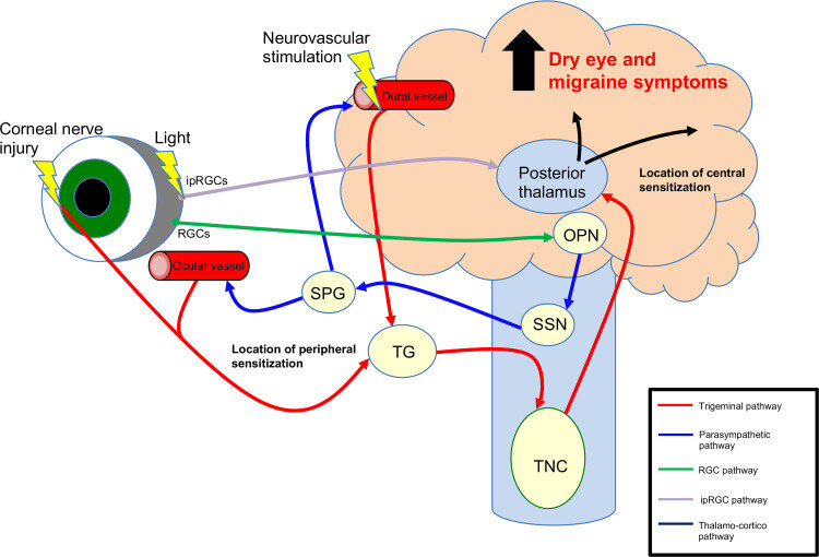
3.1 ipRGC PATHWAY (LIGHT PURPLE ARROW):
IpRGCs don’t require corneal injury to send signals, they can project directly into brain pain pathways unlike rods/cones that feed into classic vision circuits, ipRGCs send signals straight to deeper brain regions - particularly the posterior thalamus - that also receive nociceptive (pain) input from the trigeminal system. This means that light hitting the retina can activate ipRGCs -> those signals go to pain-processing nuclei in the thalamus -> those same thalamic neurons also receive trigeminal pain signals (from cornea, meninges, blood vessels) -> then the brain interprets this combined activity as photophobia/pain.
3.2 PARASYMPATHETIC PATHWAY (BLUE ARROW):
Ever happened that your eyes get red just by looking at screen? This happens to me, although it's a bit speculative, as it could technically be dry eye? But otherwise one possible mechanism is that when ipRGCs get activated by blue light, they also activate a reflex pathway in the brain. The signal travels through brain centers that control autonomic functions and ultimately activates parasympathetic nerves. This leads to dilation of blood vessels in and around the eye and even in the meninges (dural vessels in the photo). Those vascular changes can then stimulate trigeminal pain fibers, which the brain interprets as pain or photophobia.
3.3 TRIGEMINAL PATHWAY (RED ARROW):
This is the main sensory/pain pathway. When corneal nerves are already sensitized (like in CN), those two streams (retinal ipRGC signals + corneal nociceptive input), converge centrally in posterior thalamus and amplify each other, making light feel painfully sharp or deeper. The pain can be referred, in my case, I would also get pain in the V3 of the trigeminal nerve, when on computer. In the image you can see Trigeminal pathway showing in red, showing corneal nerve injury -> TG (trigeminal ganglion) -> TNC (trigeminal nucleus caudalis) -> posterior thalamus. TNC is often hyperexcitable in CN.
4. POSSIBLE LIGHT SENSITIVITY HELP?
Duloxetine lowers TNC gain, and that in theory means duloxetine could help with the light sensitivity, by modulating central amplification. I want to say, that I do in fact feel less pain (but not zero), since I'm on duloxetine (60mg). But since this is a big claim, I want to be extremely careful here, not to say this has for sure happened because of this drug. There are other drugs that also affect TNC, such as oxcarbazepine and ketamine. However they could be less effective, since duloxetine's difference is that it enhances descending inhibition from the brainstem (locus coeruleus, raphe) directly onto the TNC, including circuits processing convergent light input. That’s why duloxetine can reduce light sensitivity even if oxcarbazepine or ketamine fail - it’s "top-down" modulation instead of "local dampening". Please take this last section with a grain of salt!
Sources:
- [1] Blue light induces corneal epithelial damage by activating the NLRP3 inflammasome pathway
- [2] Exploring the Link Between Dry Eye and Migraine: From Eye to Brain
- [3] Blue light impairs cornea and corneal wound healing by downregulating VCAM1 partly
[1] After a talk with a doctor about light sensitivity, who specializes with corneal neuropathic pain, I heard there are experimental findings suggesting that under certain conditions, corneal nerves may express photopigment-like molecules. However, these have not been shown to function as true light sensors, and their role in human photophobia remains unclear.